Catalog No. CLT0002
Product information
WOS-Cy7Gal provides a rapid, simple and sensitive fluorogenic response to detect beta-galactosidase (β-gal) activity, especially the senescence associated β-galactosidase (SA-β-gal), which has been widely used as a marker of cellular senescence.
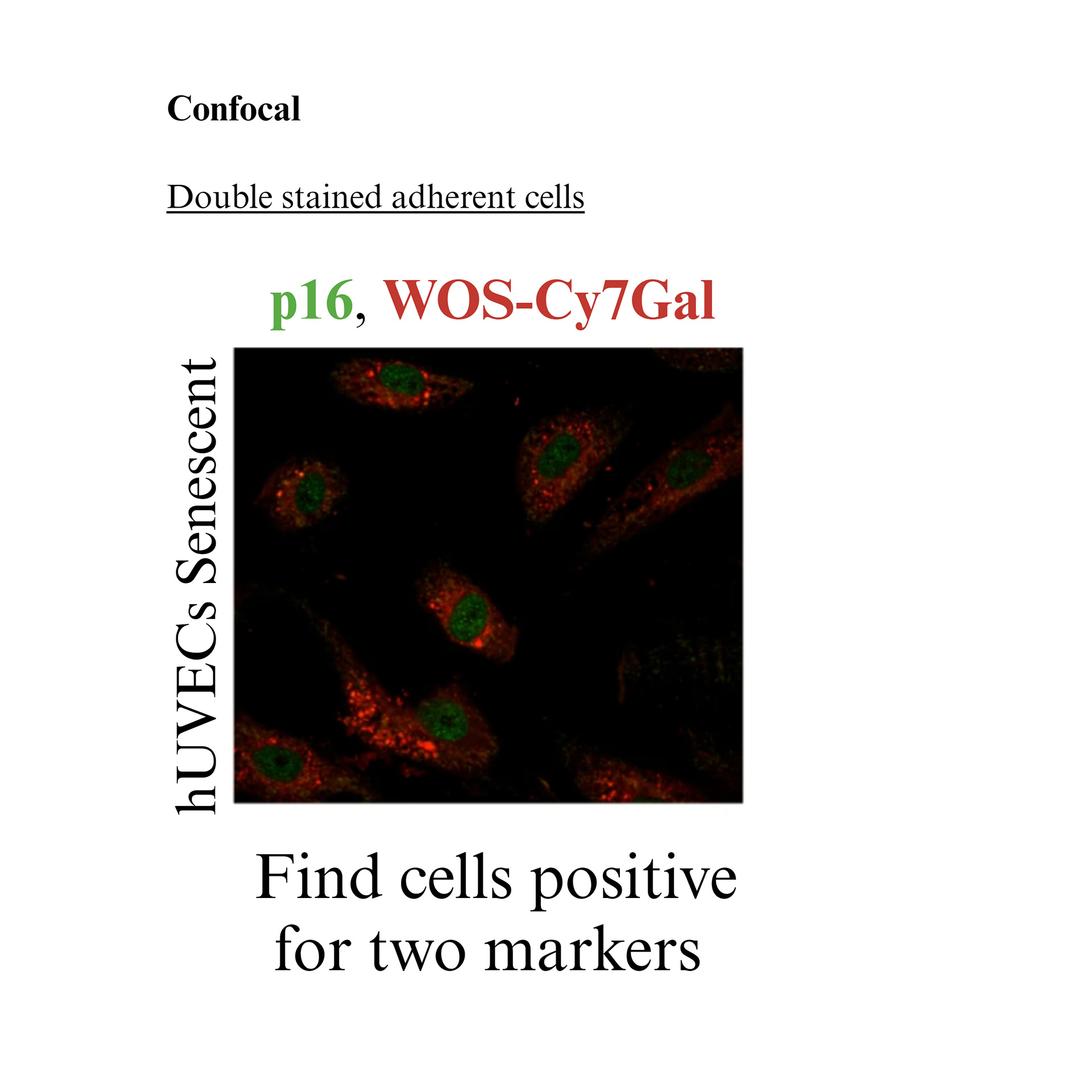
Compared to its relatives (i.e. SPiDER-bGal, Xite™ Red beta-D-galactopyranoside or resorufin beta-D-galactopyranoside), WOS-Cy7Gal produces a faster response (10-15 min) and has better retention inside the cells. Importantly its signal supports posterior fixation, permeabilization and antibody incubation protocols, making it easy to double or triple stain cells. This feature is notably valuable to unequivocally identify senescent cells, as there is no universal marker to detect them, and their explicit detection usually relies on the confirmation of the overexpression or downregulation of different proteins.
WOS-Cy7Gal probe is poorly fluorescent at 660 nm (λexc = 580 nm), releasing the highly fluorescent dye WOS-Cy7 after β-Gal hydrolysed.
Table 1. Contents and storage.
| Product | Mw (g/mol) | Catalog No. | Amount | No. of assays (12-well plate/Surface area 3.5 cm2) | Storage* |
| WOS-Cy7Gal | 876.5 | CLT0002A | 1 mg | 50 | -20°C/-80°C
Avoid freeze-thaw cycles Desiccate Protect from light |
| CLT0002B | 2 mg | 100 | |||
| * When stored as directed, the product is stable for at least 6 months at -20°C from the date receipt. | |||||
To prepare stock solutions
WOS-Cy7Gal is provided as a lyophilized powder. Prepare a 1000 X solution (20 mM) by adding 57 μL of DMSO direct to the 1 mg vial. Add 1 μL of this solution directly to 999 μL of the proper culture media. Note: WOS-Cy7Gal working solutions should be used promptly. The concentration of WOS-Cy7Gal should be optimized for different cell types and conditions.
Platform
Flow cytometry
Excitation 561 or 642 nm. From PE to APC channel
Confocal microscopy
Excitation 561 or 642 nm. From Red to Far red channel
Flow cytometry
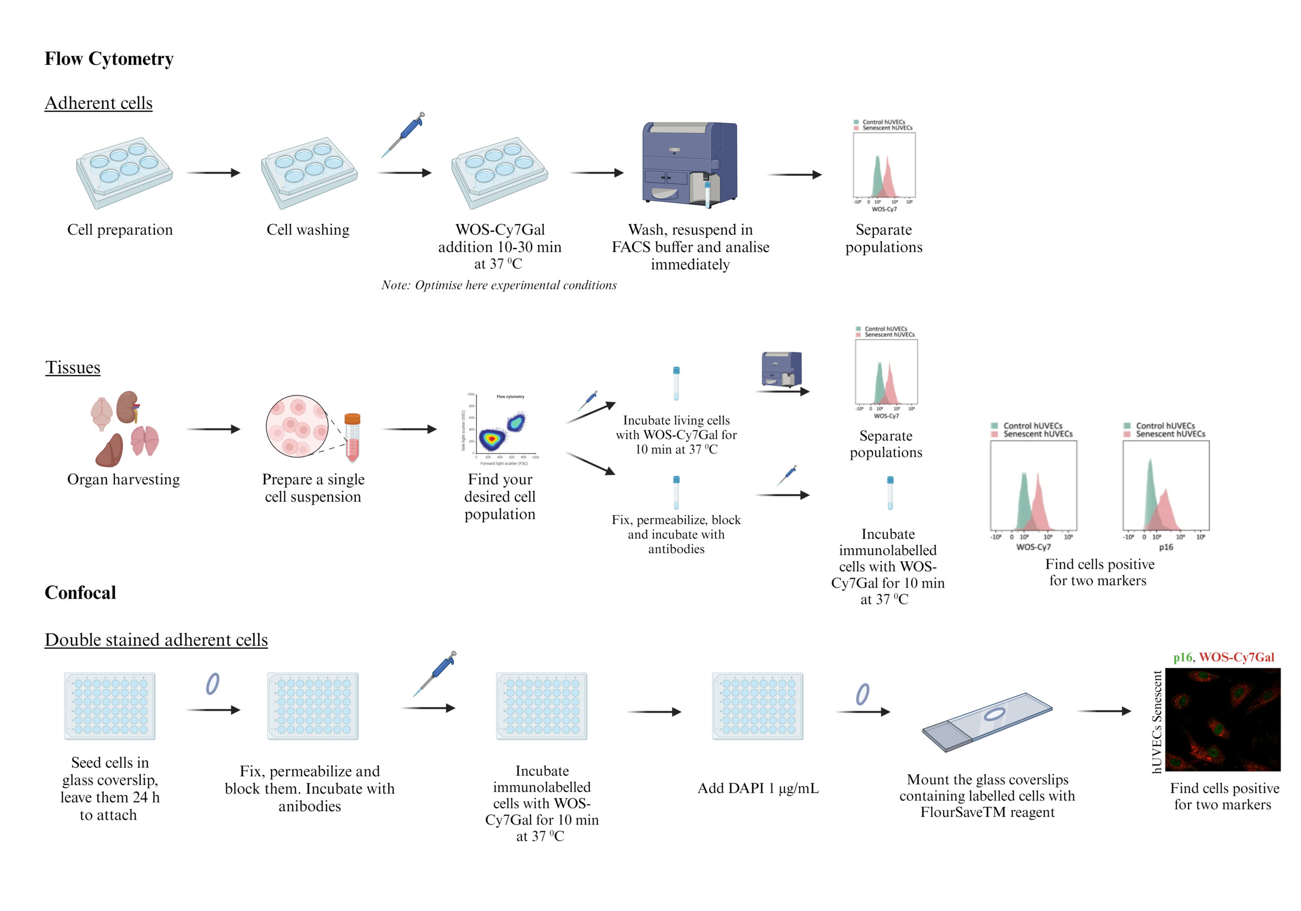
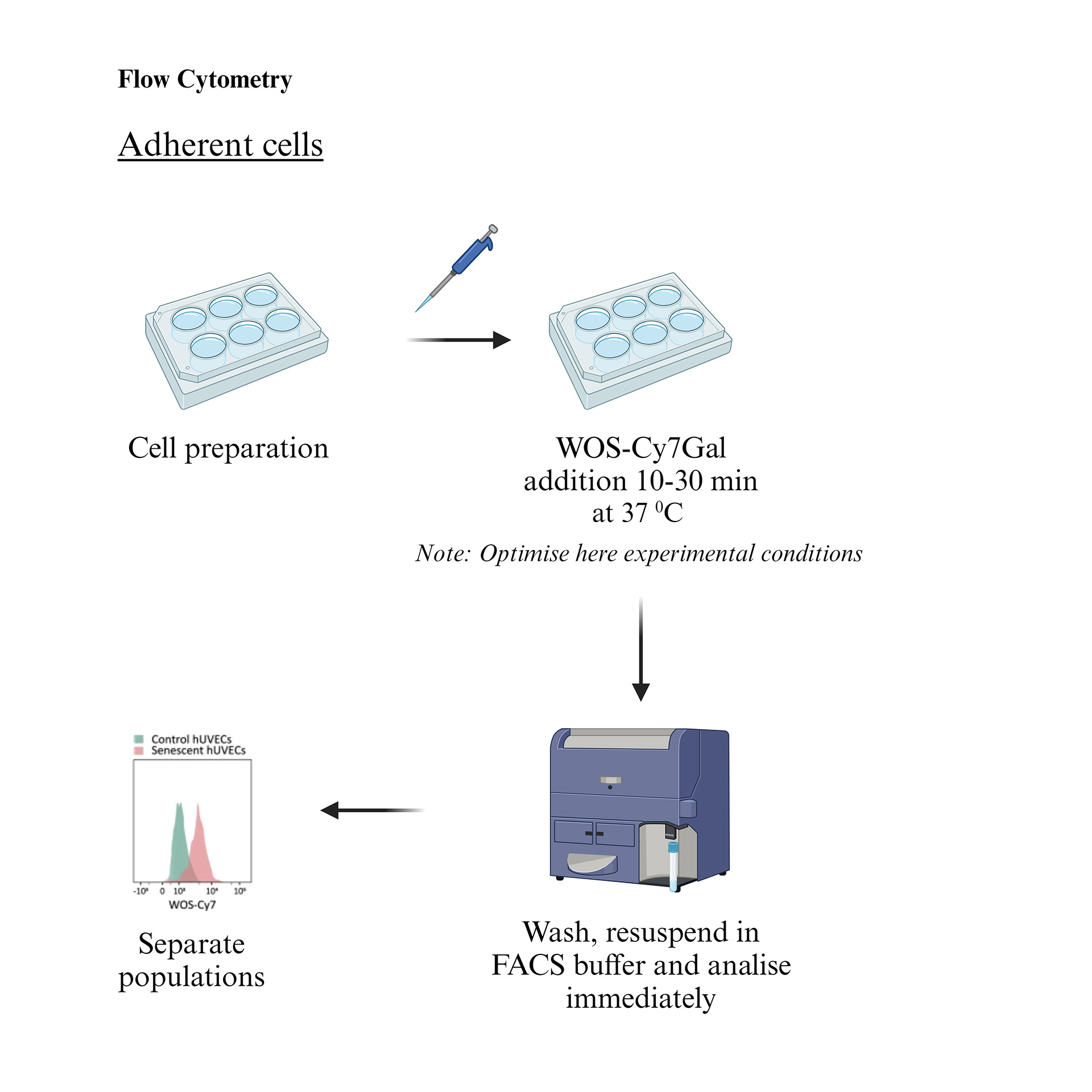
Adherent cells
- Treat your samples as desired. As an example, 50.000-80.000 cell/well can be seeded in a 12 well-plate and leave them overnight to attach.
- Remove any treatment or media and wash them with DPBS.
- Add WOS-Cy7Gal working solution for 10-30 minutes and incubate the samples at 37 °C incubator. Note: Optimal time for incubation needs to be determined experimentally.
- Remove WOS-Cy7Gal solution and wash cells with DPBS.
- Resuspend the cells in FACS buffer (10% Hank’s Balance Salt Solution, 10% EDTA, 1% HEPES, sterile H2O) and monitor the fluorescence intensity with a flow cytometer using the filter set stated above.
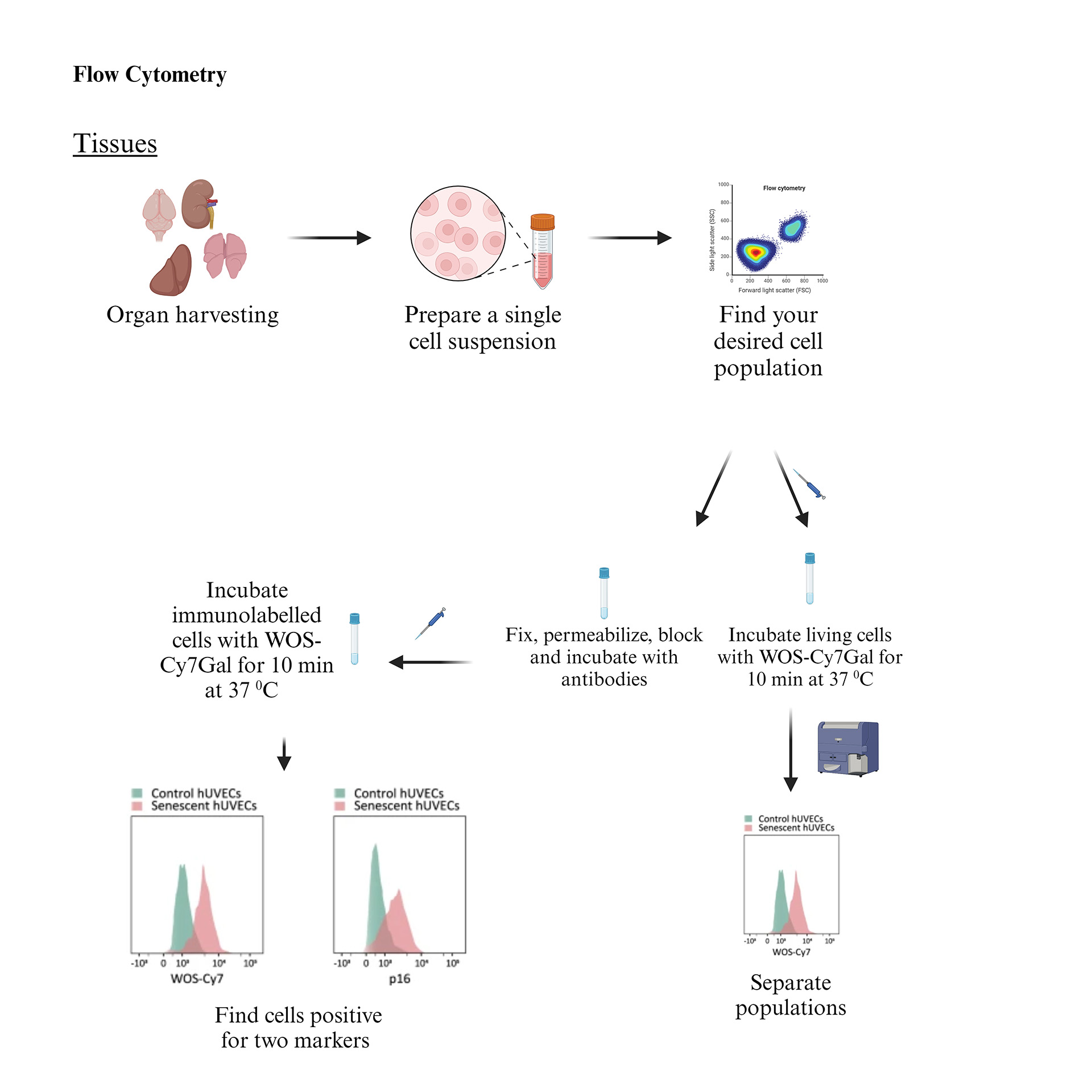
Tissues
- Harvest the desired tissue and subject it to enzymatic digestion (g. with a mixture of 1 mg/ml collagenase/dispase and 0.2 mg/ml DNAse diluted in 2 ml of culture media), dissociate the tissue mechanically and filtered through a 40 μm nylon filter to obtain a single cell suspension.
- Target your cell populations with specific antibodies to localise desired populations (g. incubate cell mixtures with CD31 and CD45 antibodies to label endothelial and immune cells respectively).
For simple SA-β-Gal detection
- Incubate live cells with WOS-Cy7Gal in FACS buffer (10% Hank’s Balance Salt Solution, 10% EDTA, 1% HEPES, sterile H2O) for 15 min prior to flow cytometry assessment. Note: It is optional to incubate the cells with 0.1 μg/ml DAPI to exclude dead cells.
For double staining, fix, permeabilize and block cells as preferred. Incubate with primary and secondary antibodies labelled with Alexa FluorTM as suggested by the manufacturer.
- Incubate with WOS-Cy7Gal working solution for 10 min.
2) Centrifuged the cells (500 xg, 5 min), resuspend them in FACS buffer, and analyse immediately.
Confocal
For simple SA-β-Gal detection
- Seed the cells in a specific chamber for optical imaging. As an example, 10.000-20.000 cell/well can be seeded in an 8 well-plate and leave them overnight to attach.
- Label the nuclei with 10 μg/ml Hoechst for 30 min.
- Wash cells carefully.
- Incubate with WOS-Cy7Gal working solution for 10 min. Note: Optimal time for incubation needs to be determined experimentally. Image the cells immediately.
For double staining
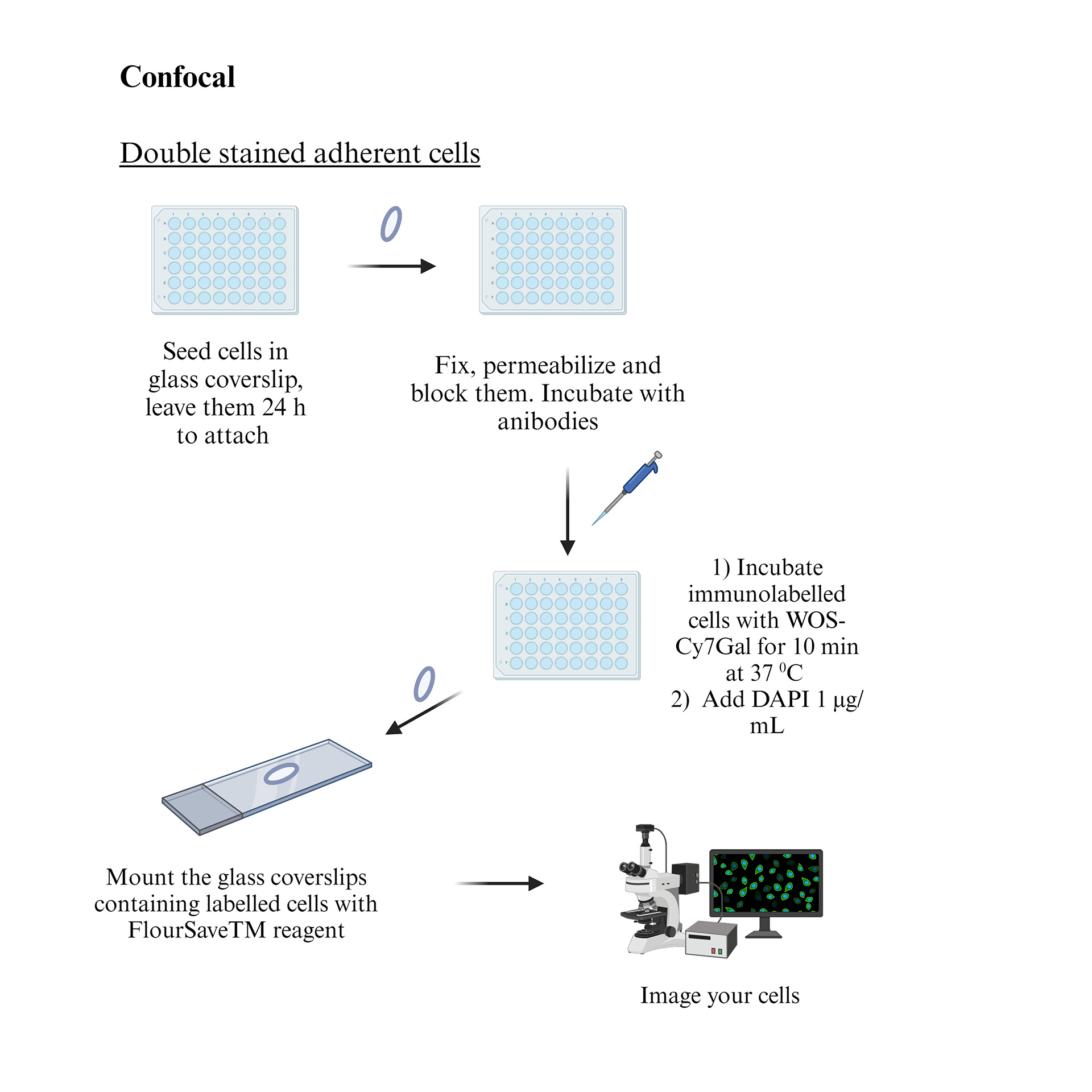
- Seed the cells in round glass coverslips. As an example, an autoclaved 1 cm2 glass coverslip can be placed in a 48-well plate and 12,500 and 25,000 cells can be plated. Leave cells to attach.
- Fix the cells as preferred (g. 2% paraformaldehyde (PFA) for 15 min and washed thoroughly with 0.1 M PBS).
- Block non-specific binding (g. 10% horse serum and 0.2% TritonTM X-100 in PBS for 1 h at RT).
- Incubated with desired primary antibodies overnight at 4 °C.
- Wash the cells carefully with DPBS.
- Incubated with desired secondary antibodies labelled with Alexa FluorTM for 1 h at RT.
- Incubate the immunostained cells with WOS-Cy7Gal working solution for 10 min at 37 °C.
- Stain nuclei with 4′,6-diamidino-2-phenylindole (DAPI, 1 μg/ml in distilled water) for 4 min and mount the glass coverslips containing labelled cells with FlourSaveTM reagent.
- Image as preferred.
- Nature Communications volume 15, Article number: 775 (2024)

Reviews
There are no reviews yet.